
Presentation
'Splayfoot' and plantar pain between the 3rd and 4th metatarsophalangeal joints.
Patient Data
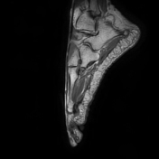

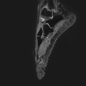
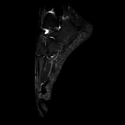
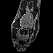


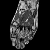
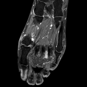
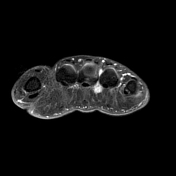

Dumbbell/ovoid-shaped lesion (~ 12 x 5 x 15 mm) in the 3rd web space between the 3rd and 4th metatarsophalangeal joints. The lesion is isointense to muscle and hypointense to the surrounding adipose tissue in T1, T2, and PD images and slightly hyperintense compared to the surrounding fat in fat-saturated PD images. It displays strong enhancement after contrast administration.
Exam courtesy: Stefanie Otto (radiographer)

Findings
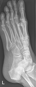
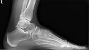
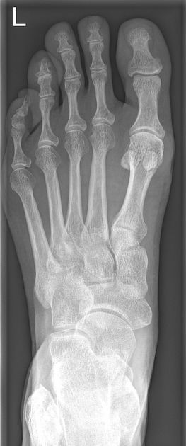
- mild degenerative changes in the 1st metatarsophalangeal joint
- first metatarsophalangeal angle: <15°
- first intermetatarsal angle: ~9° (mildly abnormal)
- fourth-fifth intermetatarsal angle: ~7°
Conclusion
- no hallux valgus, no bunionette deformity
- radiographically no signs of splayfoot
Case Discussion
Morton neuroma denotes a focal perineural fibrosis most often located in the third interdigital web space between the 3rd and 4th metacarpophalangeal joints leading to a fusiform enlargement of a common plantar digital nerve 1. Morton neuromas are a common cause of forefoot pain and on MRI they are characterized by a dumbbell-shaped contrast-enhancing lesion as in this case.
Splayfoot is an abnormal widening of the forefoot associated with flattening of the transverse arch of the foot and is associated with hallux valgus and bunionette deformity.
Radiographically it has been defined as an increase of the first intermetatarsal angle >12° and the fourth-fifth intermetatarsal angle of >8° 2 and the angles on the weight-bearing dorsoplantar foot radiograph of this case do not suggest a splayfoot deformity.
Other differential diagnoses of plantar forefoot pain include adventitial bursitis, plantar plate tear, metatarsal stress injury, or peripheral nerve sheath tumors.


 Unable to process the form. Check for errors and try again.