
Presentation
Large cervical mass in transvaginal sonography.
Patient Data
Age: 40 years
Gender: Female
From the case:
Prolapsed endometrial polyp


Download
Info

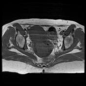
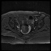
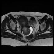
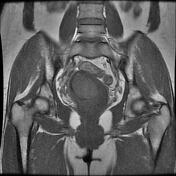
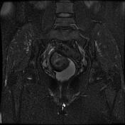
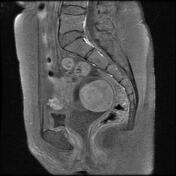
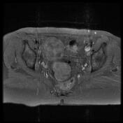
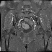
Large pedunculated intracavitary mass arising from the uterine anterior wall, filling the uterine lower segment cavity and prolapsing into the vagina through a distended endocervical canal.
The postcontrast sequences show a heterogeneous enhancement.
No pelvic lymphadenopathy is seen.
Case Discussion
MRI features are most consistent with a prolapsed endometrial polyp.


 Unable to process the form. Check for errors and try again.