
Presentation
Abdominal pain.
Patient Data
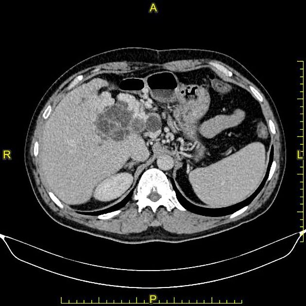
Few hepatic cysts, the largest seen at right liver lobe, which contains small intra cystic daughter cysts.
Multiple variable sized cysts seen along the course of common bile duct.
Intrahepatic biliary dilatation.
Filling defect seen within the superior mesenteric vein in keeping with non-obstructing thrombus.
Multiple collaterals in the porta hepatis area, most likely due to mass effect of the cysts on the portal confluence and portal vein.
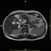

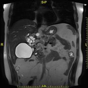
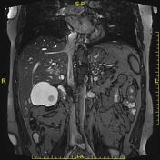
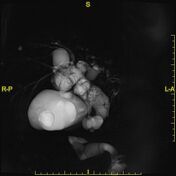


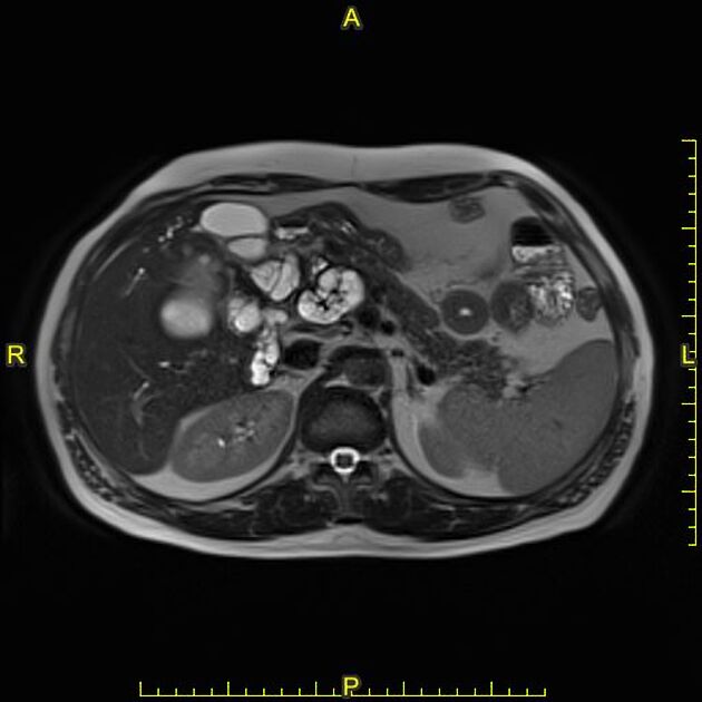
Few hepatic cysts, the largest seen at right liver lobe, which contains multiple daughters cysts.
Multiple cysts seen along the course of dilated common bile duct (intraluminal), associated with intrahepatic bile ducts dilatation.
Case Discussion
Patient is a known case of hepatic hydatid disease, underwent partial liver resection.
Radiological features are most likely in keeping with hepatic hydatic disease with intrabiliary rupture of hepatic hydatid cyst.


 Unable to process the form. Check for errors and try again.