
Presentation
Reduced vision in the right eye, ophthalmoplegia, and multiple cranial nerve palsies.
Patient Data
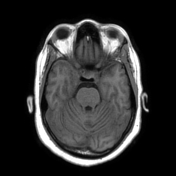

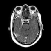
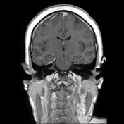
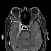

Asymmetric enlargement of the right cavernous sinus, the right cavernous sinus measures 15 mm in its widest diameter while the left measures 4 mm. Marked thickening and enhancement of the right cavernous dura with extension into the orbital apex, superior orbital fissure, and anterior temporal lobe. Associated narrowing of the cavernous part of the right ICA.
Mild right proptosis is noted.
No aneurysm, cavernous sinus thrombosis, or caroticocavernous fistula is seen. The pituitary gland, left cavernous sinus, and brain parenchyma appears unremarkable.
Case Discussion
Given the clinical presentations and MRI findings, a diagnosis of Tolosa-Hunt syndrome was made. The differential diagnosis includes sarcoidosis, inflammatory pseudotumor of the skull base, lymphoma, granulomatosis with polyangiitis, hypertrophic pachymeningitis, and lymphoma. The patient had significant improvement following steroid treatment.


 Unable to process the form. Check for errors and try again.