
Presentation
Chronic pain and movement restriction of left shoulder over six years duration.
Patient Data
CASE OF THE MONTH: This case was selected as the Case of the Month for March 2023.
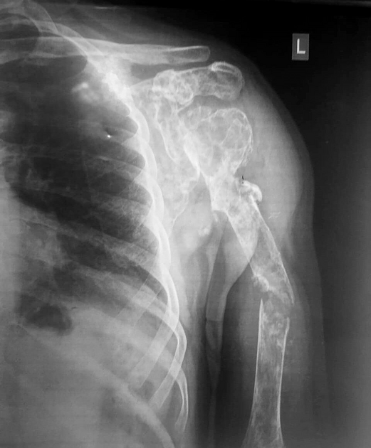
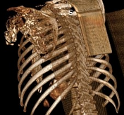
There is diffuse multiloculated expansile osteolytic lesions involving the left humerus and scapula with associated thinning of the cortex. There is a complete oblique fracture of the mid humeral shaft. There is no obvious surrounding soft tissue swelling.
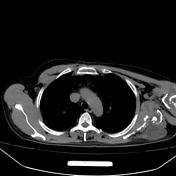

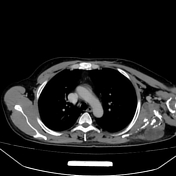
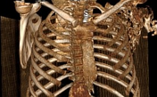

Severe bony destruction of the left scapula and proximal humerus by a multiloculated expansile cystic mass that extends to involve subscapularis, supraspinatus, infraspinatus, latissmus dorsi and deltoid muscles with density ranging from 3-17 HU, multiple calcific specks are seen within.

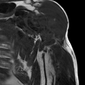
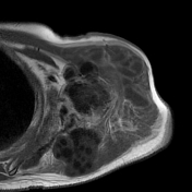
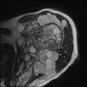
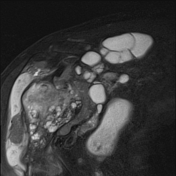
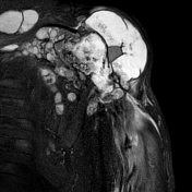
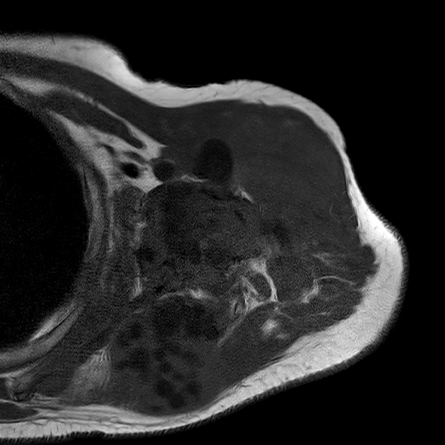
Multiloculated cystic mass composed of numerous cysts of variable size, elicits low signal intensity on T1, high signal intensity on T2 and STIR with low-intensity rim, peripherally enhanced after IV contrast, and containing multiple rounded daughter cysts.
Surgical pathology report
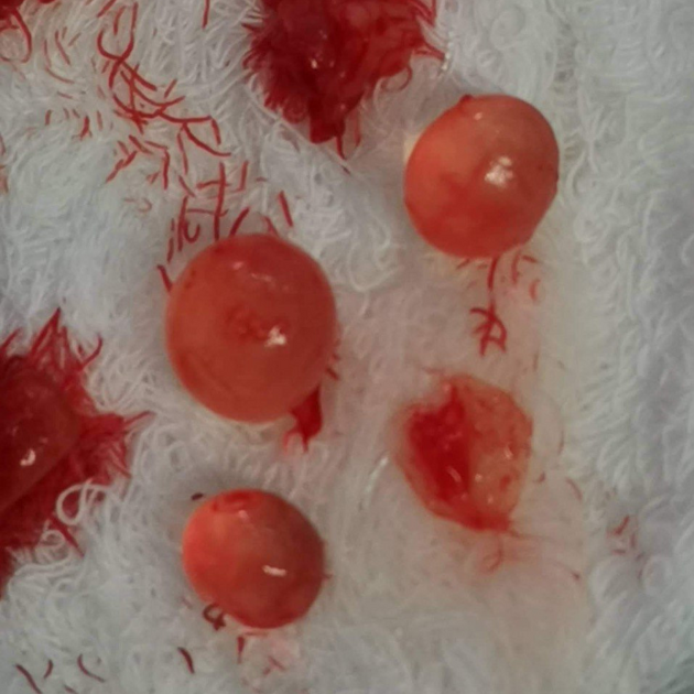
Gross:
1- Multiple bone fragments and brown soft tissue measured 3.5x3x2cm, admixed with an oval smooth cyst of the gelatin-like wall measured 2x1x1cm (block1).
2- Multiple brown soft tissues measured 4x2x2cm, including an opened cyst of the gelatin-like wall measured 2x1x1cm (block2&3).
Diagnosis;
Sections from both specimens revealed fibromuscular tissue and bone fragments involved by fibrosis and sclerosis and admixed with cyst and cyst wall pieces of lamellated eosinophilic membrane.
Features are those of hydatid cysts with reactive changes.
No malignancy was seen.
Case Discussion
Musculoskeletal hydatid infections are a very rare form of hydatid disease.
Thanks to orthopedic surgeon Dr Ali Abdulrazzaq.


 Unable to process the form. Check for errors and try again.