
Presentation
One week history of decreased cognitive and memory function, including difficulty forming new memories, acalculia and finger agnosia.
Patient Data
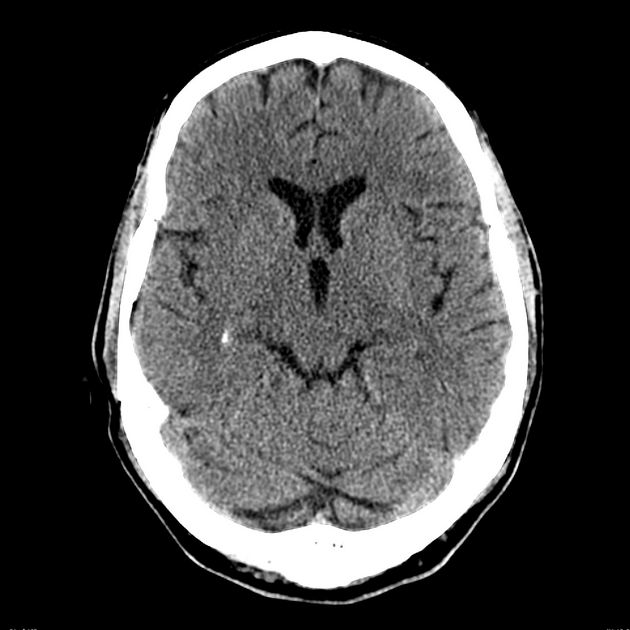
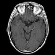
Non-contrast CT of the brain demonstrates no focal abnormality. No evidence of a stroke or tumor. No hemorrhage.
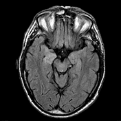

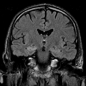
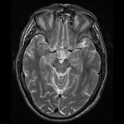
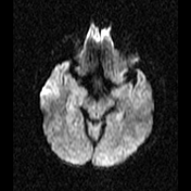

Bilateral mesial temporal lobe increased T2 signal is demonstrated, more marked on the right. It involves the amygdala and parahippocampal gyrus and extends along the hippocampal head, body and tail.
There is no convincing contrast enhancement. No evidence of hemorrhage. MR spectroscopy demonstrates minor elevation of the choline peak (not shown).
CSF was sent for extensive analysis.
- Viral PCR: negative
- NMDA Receptor Antibodies: negative
- Antinuclear Antibodie: Negative
- Extractable Nuclear Antigens: negative
- Anti - Sm: Negative
- Anti - RNP : Negative
- Anti - Ro : Negative
- Anti - La : Negative
- Anti - Topo1: Negative
- Anti - Jo1: Negative
- Anti-Endomysium: Negative
- Transglutaminase IgA: Negative
- Anti Hu: Negative
- Anti Ri: Negative
- Anti Yo: Negative
- Anti VGKC Antibody: detected and elevated
Case Discussion
This is a case of autoimmune limbic encephalitis. When an auto-antibody is found it is usually to either VGKC or NMDAR.
Antibodies to the voltage-gated potassium (VGKC) channel interfere with peripheral neuromuscular and ganglionic transmission and may be associated with a number of neurological syndromes, including limbic encephalitis, neuromyotonia and seizures. Anti-VGKC antibodies are tested by radioimmunoassay.
NMDAR antibody (negative in this case) was first found in young female patients with ovarian tumors and prominent psychiatric symptoms, amnesia, seizures, dyskinesias, autonomic dysfunction and decreased levels of consciousness. The antibodies are now also found in males and females with no known tumor and in children. Their presence suggests an immunotherapy-responsive condition 1.


 Unable to process the form. Check for errors and try again.