
Presentation
Sciatica.
Patient Data


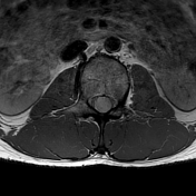
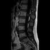
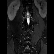
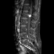
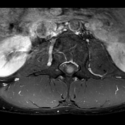
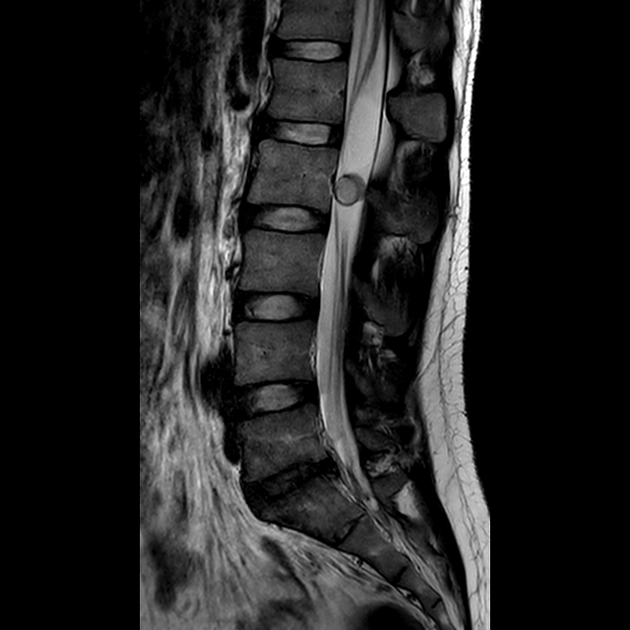
A well-circumscribed mass below the conus has fairly high T2 signal, intermediate T1 signal and vivid contrast enhancement. No flow voids. No leptomeningeal nodularity or deposits. It is a solitary lesion.
The differential includes myxopapillary ependymoma, paraganglioma or schwannoma.
Case Discussion
The patient went on to have a resection.
Histology
The sections show a well-circumscribed tumor composed of nests and cords of tumor cells amongst a prominent vascular network. The tumor cells are monomorphic with round-oval nuclei and fine-speckled chromatin, and most have an epithelioid appearance. There are vague perivascular pseudo-rosettes. There is no myxoid material or papillary architecture.
By immunohistochemistry, the tumor cells are positive for synaptophysin and chromogranin A, and negative for EMA and GFAP. 5100 and SOX10 highlight scattered single cells. The tumor cells show intact staining for SDHA and SDHB indicating that SDHX mutation is unlikely.
The lesion appears to have been shelled out with very narrow (<0.1mm) but clear margins in the planes examined.
Final diagnosis
paraganglioma of the filum terminale (WHO grade 1) - now known as a cauda equina neuroendocrine tumor.


 Unable to process the form. Check for errors and try again.