
Presentation
Pain right hypochondrium associated with nausea. No urinary symptoms.
Patient Data
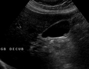
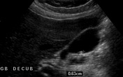
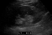
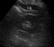
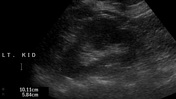
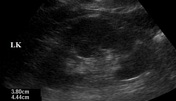
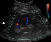
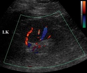

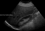
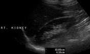
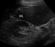
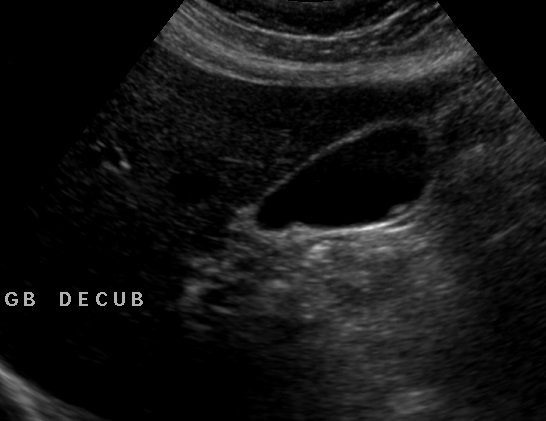
Two small gallstones without any sonographic features of acute cholecystitis are noted. A focal bulge or prominent hump is seen at the mid pole of the left kidney which has the echogenicity similar to the adjacent cortex. Vessels are seen coursing through it normally without any mass effect or distortion. Right kidney is normal.
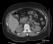

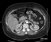

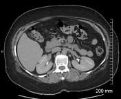

No suspicious focal mass lesion is seen in the left kidney. A simple renal cyst measuring 7 x 9 mm is seen in the right kidney. Prominent bilateral extrarenal pelvis (anatomical variant). Uterine leiomyoma measuring 3.5 x 4.0 cm having central non-enhancing cystic area which is likely necrosis.
Case Discussion
Dromedary hump is a normal variation in the renal contour caused by the splenic impression along the superolateral aspect of the left kidney and should not be mistaken for a mass lesion.
Our case has classical sonographic features of a dromedary hump; however, unfortunately, it was misinterpreted as a renal mass which prompted further investigation with CT urography.


 Unable to process the form. Check for errors and try again.