
Presentation
Long standing epilepsy. Started in right hand.
Patient Data
Age: 40 years
Gender: Female
From the case:
Polymicrogyria


Download
Info

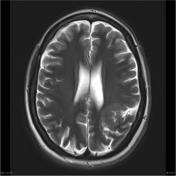
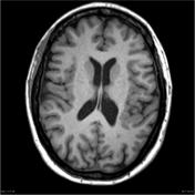
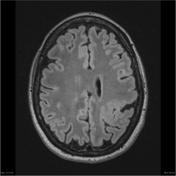
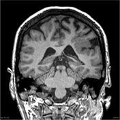
MRI of the brain demonstrates an extensive area of developmental malformation which involves the posterosuperior margins of the left Sylvan fissure. This is characterized by abnormal cortical thickening and a nodular surface and grey-white matter interface. No cleft or grey matter heterotopia noted. Septum pellucidum is present.
Case Discussion
This case demonstrates typical appearance of polymicrogyria which in this case is unilateral.


 Unable to process the form. Check for errors and try again.