
Presentation
Unconscious collapse lasting 30 seconds.
Patient Data
Age: 20 years
Gender: Male
Download
Info

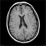
Non contrast CT demonstrates a nodule of tissue bulging into the left lateral ventricle with density similar to grey matter.


Download
Info
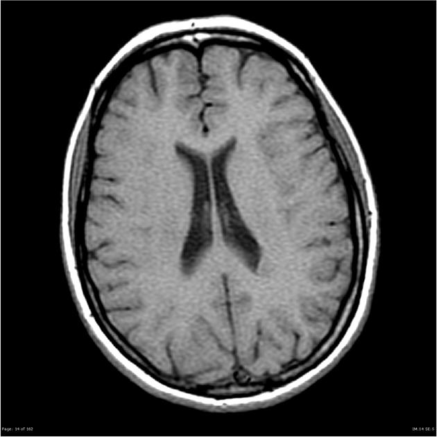
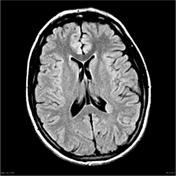
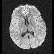
MRI demonstrates a 15 x 8 x 10 mm nodule which projects into the trigone of the left lateral ventricle. It has signal characteristics which match those of cortex on all sequences and demonstrates no abnormal contrast enhancement. There is no convincing abnormality of the overlying cortex.
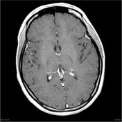
Incidental note is made of a small pineal cyst which measures 10 mm in maximal diameter. No abnormal contrast enhancement.
Case Discussion
CT and MRI demonstrate typical appearances of an isolated and sporadic nodule of periventricular grey matter heterotopia.


 Unable to process the form. Check for errors and try again.