
Presentation
Acute on chronic left 2nd-digit pain and discomfort. Negative recent trauma.
Patient Data

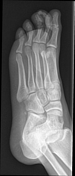

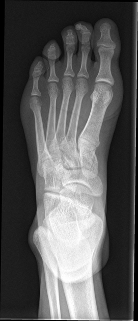
Plain films demonstrate a subungual exostosis of the left second digit. There are no aggressive features or a "bizarre" appearance. The left foot is otherwise normal.

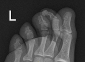
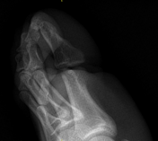
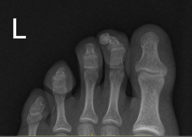
Zoomed images of the left second digit demonstrate the exostosis much better. The exostosis is typically subungual in anatomical location and appears attached or continuous with the terminal phalanx. It is well-corticated and eccentric towards the lateral margin of the terminal phalanx.
Case Discussion
The appearance, anatomical location within the subungual space (nailbed) and attachment to the terminal phalanx are highly suggestive of a subungual exostosis.
The differential diagnosis includes a turret exostosis and a bizarre parosteal osteochondromatous proliferation (BPOP). Both these lesions are less likely due to the above-mentioned typical features.


 Unable to process the form. Check for errors and try again.