
Presentation
NA
Patient Data
Age: Adult
From the case:
Adrenal glands (normal CT anatomy)

Show annotations
Download
Info

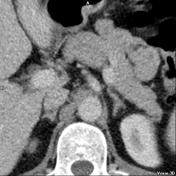
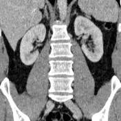
Axial and coronal cropped CT focusing on normal appearance of adrenal glands on CT. On the axial CT the right adrenal gland is seen posterior to the inferior vena cava, while the left adrenal gland is seen posterior to the pancreas. On the coronal CT the right adrenal gland is seen medial to the right lobe of the liver and lateral to the right diaphragmatic crus, while the left adrenal gland is seen medial to the pancreatic tail/spleen and lateral to the left diaphragmatic crus.
From the case:
Adrenal glands (normal CT anatomy)
Download
Info

Yellow arrows on axial image indicate the adrenal glands.
Case Discussion
Annotated zoomed CT images of the adrenal glands.


 Unable to process the form. Check for errors and try again.