
Presentation
Short stature.
Patient Data
Age: Child
From the case:
Ectopic posterior pituitary
Download
Info
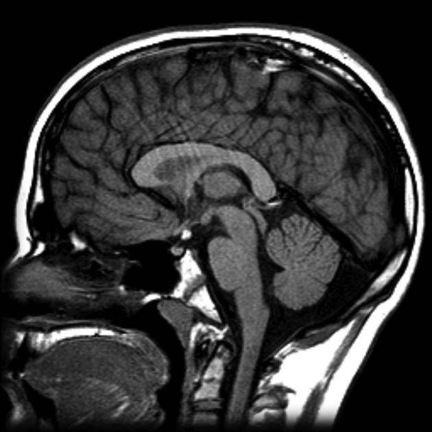
Single midline sagittal T1 image demonstrates a nodule of intrinsic high T1 signal attached to floor of the third ventricle. No normal posterior pituitary is visible in the fossa.
Download
Info
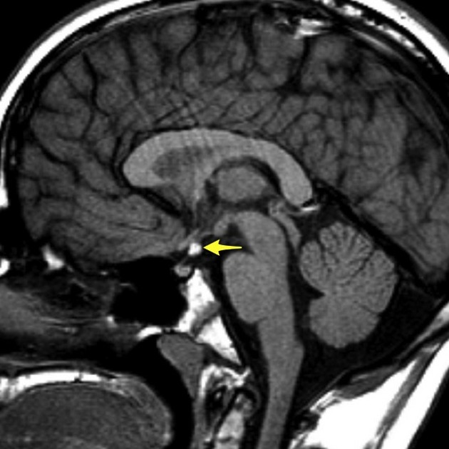
No normal posterior pituitary bright spot is seen in the pituitary fossa, but rather a small region of high T1 signal (yellow arrow) is seen high up at the top of the infundibulum (which appears small).
Case Discussion
This case illustrates typical appearances of an ectopic posterior pituitary with associated growth hormone deficiency.


 Unable to process the form. Check for errors and try again.