
Presentation
Presented to ER with hypoglycemic coma. Arrested twice.
Patient Data
Age: 25 years
Gender: Female
From the case:
Extensive hypoxic-ischemic insult


Download
Info
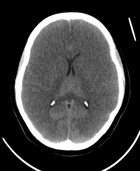
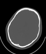
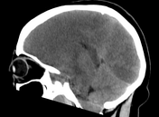
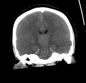
Diffuse cerebral edema and low attenuation with loss of grey-white matter interface, compression of the lateral ventricles and effacement of the cerebral cisterns and extra-axial CSF spaces. The cerebellum is spared (white cerebellum sign). Also, there's cerebellar tonsil herniation.
Case Discussion
In this case, CT demonstrates extensive ischemic damage (e.g.cerebral edema, reversal sign, and herniation), which are suggestive of brain death. However, only CT/MRI angiography is recognized as a potential alternative to angiography and nuclear medicine studies in diagnosis of brain death.
CT angiography relies upon the non-opacification of intracranial vessels.


 Unable to process the form. Check for errors and try again.