
Presentation
Sciatica.
Patient Data
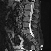

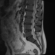


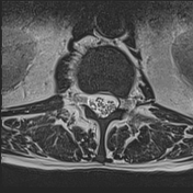
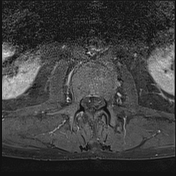
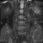

A lesion within the thecal sac intimately associated with the cauda equina at the level of L2/L3 has vividly enhancement and prominent flow voids superiorly. It has quite high T2 signal.
The bone marrow signal is low similar but not lower, than muscle. on T1. Diverticular change of the colon noted.
Conclusion
Cauda equina mass with prominent flow voids favors a cauda equina neuroendocrine tumor, over the more common differential diagnoses of myxopapillary ependymoma and schwannoma.
Bone marrow signal suggests florid red marrow (e.g. anemia) or infiltration (e.g. leukemia).
Case Discussion
The patient went on to have a resection.
Histology
Sections show a well-circumscribed partially encapsulated tumor composed of nests of cells with abundant eosinophilic cytoplasm, uniform cells with a granular salt and pepper chromatin separated by richly vascular fibrovascular septae. Areas of ganglion cell metaplasia and schwannian stroma are present.
The tumor is positive for INSM1 and synaptophysin, and focally chromogranin. S100 shows focal presence of sustentacular cells surrounding nests. There is weak positivity far GFAP. The tumor is negative for AE1/AE3, EMA and GATA3.
Final diagnosis
Cauda equina neuroendocrine tumor (paraganglioma)
They also had a full blood examination that confirmed the presence of severe normocytic and normochromic anemia accounting for the bone marrow abnormality. This was found to be due to bowel blood loss from small bowel angiodysplasia.
Hb = 79 g/L
Hematocrit 0.24 L/L (N 0.36 to 0.50)
High ferritin = 582 ug/L


 Unable to process the form. Check for errors and try again.