
Presentation
No neurological symptoms. MRI brain done with MRI of the spine as a work up for scoliosis.
Patient Data
Age: 8 years
Gender: Female
From the case:
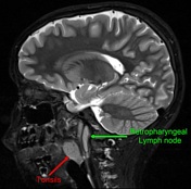
Retropharyngeal lymph nodes


Download
Info
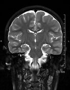
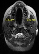
Small bilateral retropharyngeal lymph nodes at the skull base. Multiple small sub-centimeter lymph nodes, likely reactive in nature, are seen on both sides of the neck. Enlarged palatine tonsils. No significant nasopharyngeal adenoid enlargement is noted.
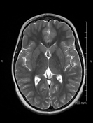
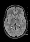
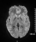
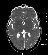
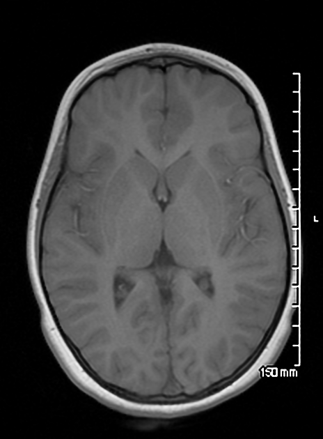
No hemorrhage or infarction was identified. No focal mass lesion, hydrocephalus, mass effect or midline shift. Cerebellum and brain stem are normal. Basal cisterns are normal.
Download
Info
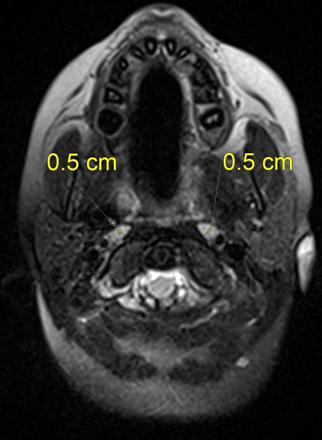
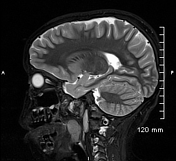
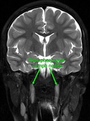
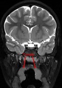
Small sub-centimeter bilateral retropharyngeal lymph nodes and palatine tonsils.
Case Discussion
Incidental finding of small bilateral retropharyngeal lymph nodes at C1/C2 level (lateral group).


 Unable to process the form. Check for errors and try again.