
Presentation
6 months of gradual worsening left sided vision, headache and weight gain.
Patient Data
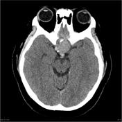

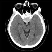
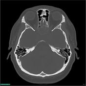

CT demonstrates a large mass centered on the central base of skull, in the region of the pituitary fossa. A slightly hyperdense component extends superiorly indenting the floor of the third ventricle. The mass demonstrates moderate enhancement and encases both carotid arteries. The bone is remodeled and thinned with areas appearing deficient inferiorly.
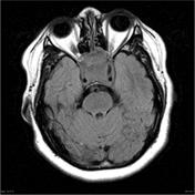
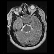

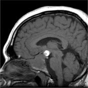
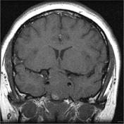
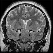

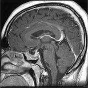
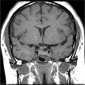
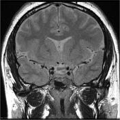
A large predominantly solid multilobulated mass measuring 3.6 x 3.8 x 4.1cm is centered in an expanded sella, with no separate pituitary gland identified. The mass is predominantly isointense to cerebral parenchyma on T1 and T2 weighted imaging and heterogenously contrast enhancing. No diffusion restriction. It extends into sphenoid and posterior ethmoid sinuses, and the suprasellar and interpeduncular cisterns, compressing and displacing superiorly the right aspect of the optic chiasm and surrounds the cavernous carotids within the cavernous sinuses without cavernous carotid narrowing. The soft tissue reaches the orbital apices without intraorbital invasion.
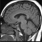
A 12mm T1 heterogeneously hyperintense, peripherally T2 hypointense component lies in the suprasellar cistern and just above the level of the tip of the basilar artery. The MRA examination demonstrates that the T1 hyperintense components is separate from the basilar tip.
No hydrocephalus although the floor of the 3rd ventricle is distorted.
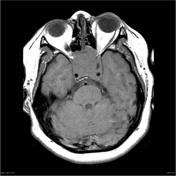
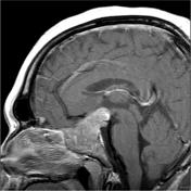
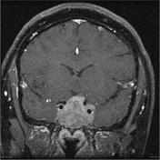
This patient was prescribed a dopamine agonis (cabergoline) and followed clinically, biochemically and with serial imaging. The following MRI was obtained 6 months after the initial scan. No surgery has been performed.


There has been remarkable decrease in the size of the pituitary macroadenoma. There is now only small amount of tissue located in the anterior part of the enlarged pituitary fossa, anterior to the normal pituitary tissue. Some tissue continues to bulge into the sphenoid sinus, but this component also has significantly reduced in size. The optic chiasm which had been elevated and compressed by the aforementioned mass has now returned to near-normal position slightly drooping into the enlarged pituitary fossa.
There is no evidence to suggest a defect in the floor of the pituitary fossa and no fluid within the sphenoid sinus to raise suspicion of a CSF leak. The remainder of the study is unremarkable.
Case Discussion
This patient was treated only with cabergoline, and demonstrates the dramatic effect it can have on prolactinomas.


 Unable to process the form. Check for errors and try again.