
Presentation
Abdominal discomfort
Patient Data
Age: 35 years
Gender: Male
From the case:
Scrotal varicocele


Show annotations
Download
Info
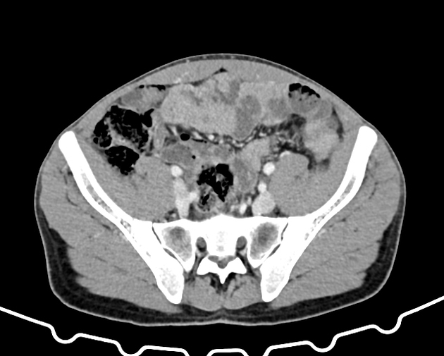
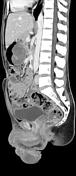
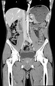
Bulky, contrast-opacified left scrotal varices. Otherwise, there were no significant findings in the abdomen or pelvis.
Case Discussion
A simple case of bulky left scrotal varices on CT is helpful to review to ensure that this is not over-called an enhancing scrotal mass. Evaluation for causes of varices can also be helpful, as CT can be used to rule out pelvic/retroperitoneal adenopathy or other pathology that may be impeding the flow of venous return through the gonadal veins.


 Unable to process the form. Check for errors and try again.