
Presentation
Right upper quadrant pain with jaundice.
Patient Data
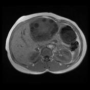

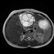
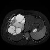
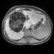
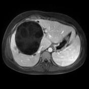
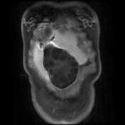

Two large well-circumscribed lobulated multiloculated hepatic cystic masses, one centered on the right lobe (mainly the segments, 4, 8, and 7) and the other one on the left lobe (mainly the falciform ligament). Both cystic masses are composed of numerous daughter cysts of various sizes mainly of peripheral location within the cystic matrix. Serpiginous linear structures of a low signal on T2 and T2 fat sat of the membranes within the mass. No enhancement was seen on the postcontrast sequences.
Mild dilatation of the intrahepatic biliary ducts (IHBD) around the cystic mass located in the right lobe with fistulous tracts between the cyst and the IHBD well-demonstrated on postcontrast sequences indicating a cystobiliary communication. No dilatation of the CBD.
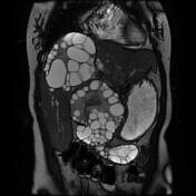
The cystic mass located in the left lobe shows subhepatic extension displacing the antropyloric region and pancreas.
Another partially calcified cystic mass is noted in the left hepatic lobe.
Case Discussion
MRI features of hepatic hydatid cysts, the largest cyst located in the right hepatic lobe shows cystobiliary communication.


 Unable to process the form. Check for errors and try again.