
Presentation
Pelvis pain for 3 weeks.
Patient Data


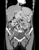
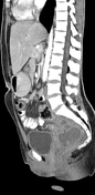
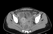
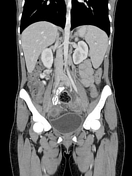

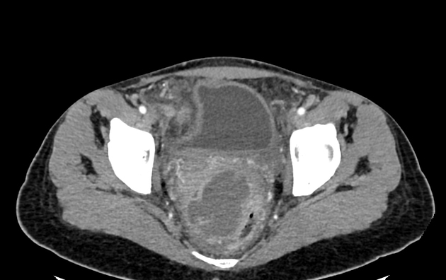
Complex multiloculated enhancing collection in the pouch of Douglas, displacing the rectum posterolaterally, with some associated reactive rectal wall thickening. Small associated collection within the left ovary. Complex rounded cystic lesion in the right ovary. Generalized stranding within the pelvis. Mild thickening of the urinary bladder. Normal appendix.
Case Discussion
Typical imaging features of an advanced tubo-ovarian abscess result in a complex, enlarged, multiloculated abscess in the pouch of Douglas as well as a smaller component of fluid in the left ovary. The cystic lesion in the right ovary may or may not be infected; it may alternatively be a hemorrhagic cyst. The picture of peritoneal inflammation of the pelvis is helpful for confirming this diagnosis and can be evidenced by bowel wall thickening and fat stranding.


 Unable to process the form. Check for errors and try again.