
Presentation
Seizure
Patient Data
Age: 10 years
Gender: Male


Show annotations
Download
Info
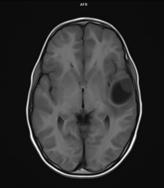
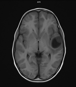
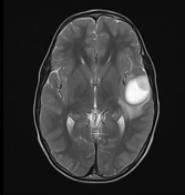
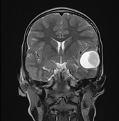
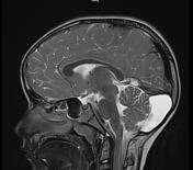
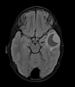
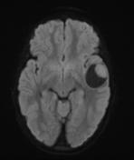
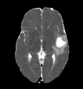
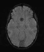
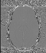
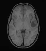
There is a well-defined solid-cystic lesion in the left temporal lobe with mild surrounding vasogenic edema. The solid component of the lesion shows vivid enhancement on post-contrast images with no evidence of calcification or hemorrhage or flow void within it. On the FLAIR sequence, cystic areas show hyperintensity relative to CSF.
Small arachnoid cyst/ Mega cisterna magna seen in the posterior fossa in the midline.
Case Discussion
The imaging findings are probably due to pleomorphic xanthoastrocytoma. It is the WHO grade 2/3 glial tumor in the young patient.
The differential diagnosis includes - ganglioglioma and pilocytic astrocytoma.
Co-author: Dr. Vivek Ranjan (Consultant pediatrician).


 Unable to process the form. Check for errors and try again.