
Presentation
Typical trigeminal neuralgia presentation on the right side.
Patient Data
Age: 30 years
Gender: Male


Download
Info
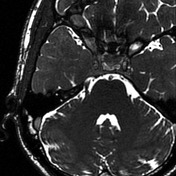
Superior cerebellar artery is seen to impress the root of trigeminal nerve, with mild buckling of the nerve.
From the case:
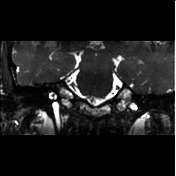
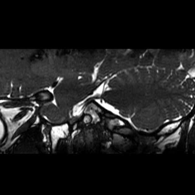
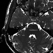
Trigeminal neuralgia: vascular compression

Download
Info

Annotated T2WI demonstrating relationship of the right superior cerebellar artery (SCA) to the trigeminal nerve.
Case Discussion
One of the common causes of trigeminal neuralgia, where MRI is particularly helpful, is compression of trigeminal nerve by loops of superior cerebellar artery or anterior inferior cerebellar artery. This case classically depicts such abnormality. Patient underwent surgery, and diagnosis was confirmed.


 Unable to process the form. Check for errors and try again.