
Presentation
7/52 headaches, inco-ordination and nausea. CT brain shows posterior fossa lesion - cystic / solid. ? hemangioblastoma for further assessment and pre op planning
Patient Data
Age: 63
Gender: Female
From the case:
Cerebellar hemangioblastoma
Download
Info
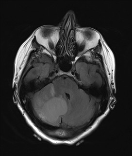
In order - T2 axial, FLAIR (sag, axial, coronal), post Gadolidium T1 axial.
Cystic lesion with fluid similar to CSF (as seen on FLAIR sequences). A solid component which is enhancing on Post GAD is seen in the periphery of the lesion.
Case Discussion
This is typical of a cerebellar hemangioblastoma.
Differential diagnosis: pilocytic astrocytoma. However will not expect this kind of enhancement on periphery.


 Unable to process the form. Check for errors and try again.