
Presentation
Memory disturbances, diplopia. A non-contrast CT revealed bilateral abnormalities. An MRI was thus performed.
Patient Data
Age: 40 years
Gender: Male


Download
Info

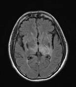
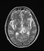
MRI revealed bilateral near symmetric abnormal signal on T2 and FLAIR with punctate hemorrhages on the region of basal ganglia bilaterally including both thalami.
The T1 sagittal images in midline reveal a thrombus in the straight sinus and internal cerebral veins. The venous thrombosis is also appreciated in Gradient echo axial images.
Case Discussion
The occurrence of deep venous thrombosis involving the straight sinus and the internal cerebral veins is uncommon. Bilateral symmetric basal ganglia/ thalamic hyperintensities should raise a suspicion of this entity and MR venography need to be added.


 Unable to process the form. Check for errors and try again.