
Presentation
Incidentally detected following a left shoulder injury.
Patient Data
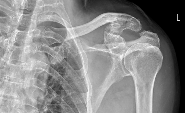
The conoid tubercle of the clavicle is enlarged and elongated, with its inferior surface approaching the superior surface of the coracoid process of the scapula to form a synovial joint (coracoclavicular joint). Degenerative changes in this joint are also observed, such as osteophytes, subchondral sclerosis of the articular surface, and irregular joint surfaces.
No dislocation of the glenohumeral or acromioclavicular joints is noted. No fractures of the shoulder region are identified.
Case Discussion
The imaging findings are consistent with a coracoclavicular joint with degenerative changes.
This is a normal anatomical variant of the pectoral girdle, which may be unilateral or bilateral. The coracoclavicular joint is a true synovial joint, usually asymptomatic and incidentally detected. Clinical symptoms arise when degenerative changes lead to osteoarthritis.


 Unable to process the form. Check for errors and try again.