
Presentation
Progressive minor weakness, particularly in the right arm.
Patient Data
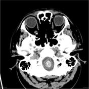

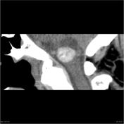
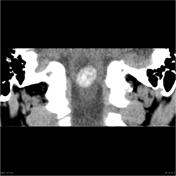

Non-contrast CT brain demonstrates a lesion within the medulla. The solid component more anteroinferiorly is densely hyperdense. There is a cystic component posterosuperior to this. There are no fatty elements identified within the lesion.
The patient was observed over a number of years, with minor but progressive symptoms. The provisional diagnosis had been of an atypical cavernous malformation, or of an unusual intramedullary tumor. Eventually the decision was made to remove the lesion as slow growth was confirmed.

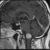
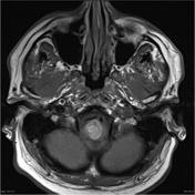
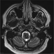
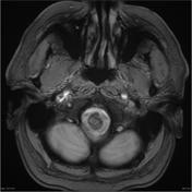
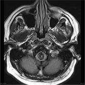
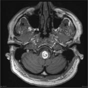
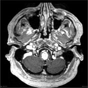

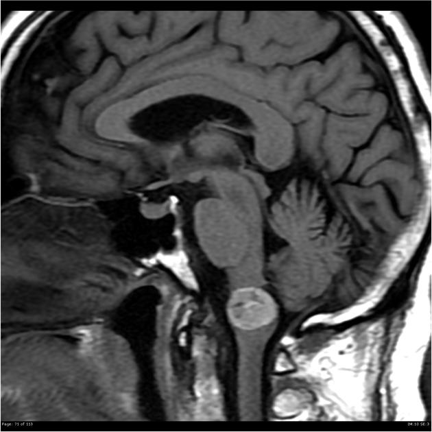
A heterogeneous inferior medullary mass is noted that is strikingly T1 hyperintense and mostly T2 hypointense with moderate magnetic susceptibility, in keeping with calcification and/or hemosiderin/melanin, with no convincing enhancement or diffusion restriction. Of note, the eccentric medullary cyst, centered over the right posterosuperior margin of the mass, has decreased in size, with minor adjacent marginal parenchymal T2 hyperintense signal abnormality.
MICROSCOPIC DESCRIPTION: The sections show a moderately hypercellular tumor. This consists of epithelioid and spindle cells which are heavily laden with globular brown pigment and show strong immunostaining for the melanocyte markers, tyrosinase and melan A. Tumor cells show moderate nuclear pleomophism with vesicular nuclei containing conspicuous nucleoli. No mitotic figures are identified and the topoisomerase index is <1%. A small focus of coagulative necrosis is also noted. Focal dural attachment is identified in one tumor fragment.
DIAGNOSIS: Melanocytic tumor with features favoring meningeal melanocytoma.
Case Discussion
Meningeal melanocytomas are rare tumors, with can be seen anywhere along the neuraxis, but have a predilection for the foramen magnum. The intense T1 hyperintensity and slow progression (compared to melanoma metastasis) are typical.
Histology courtesy of Dr M Gonzales, Royal Melbourne Hospital, Australia


 Unable to process the form. Check for errors and try again.