
Presentation
Pain with left thumb movements for the last few weeks. No trauma.
Patient Data
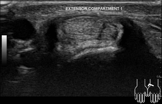
No retinacular thickening of the 1st extensor compartment. No tenosynovitis.
1st CMC joint shows capsular thickening, marginal osteophytes, ossified body, and capsular mild hypervascularity. Intact flexor capri radialis tendon.
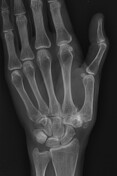
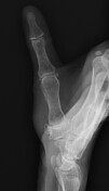

1st CMC joint shows space reduction, subchondral sclerosis, marginal osteophytes, and ossified body. No bone lesion/ periosteal reaction/ acute injury.
Case Discussion
The patient with thumb pain was referred for an ultrasound to check for 1st extensor compartment tenosynovitis. Ultrasound examination was negative for the De Quervain tenosynovitis. However, there were ultrasound findings of 1st CMC degenerative joint disease for which radiographs were taken later.
Before holding the probe, there were two red flags against the tenosynovitis diagnosis. First, the site of pain at the 1st CMC location, but not the distal radius location. Secondly, there was a visible volar side bulge at the 1st CMC region.


 Unable to process the form. Check for errors and try again.