
Presentation
Abdominal pain with palpable pelvic mass.
Patient Data
Age: 55 years
Gender: Female
From the case:
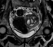
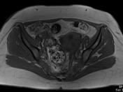
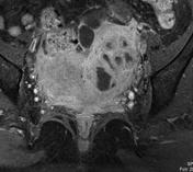
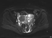
Tubo-ovarian abscess


Download
Info
Ultrasound performed by gynecologist prior to MRI request - query degenerative fibroid
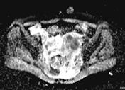
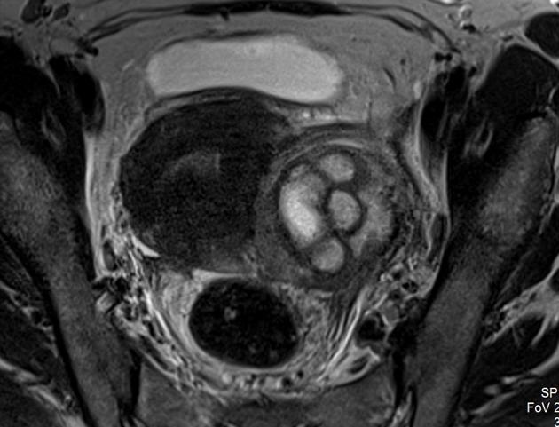
Multi-septated thick walled mass on the left side of the pelvis, displacing the uterus medially. This contains focal cystic filled spaces, which exhibit diffusion restriction. The wall and septa of the mass avidly enhance. Perilesional free fluid. Ovary not separately identified.


 Unable to process the form. Check for errors and try again.