
Presentation
Ankle pain
Patient Data
Age: 35 years
Gender: Male
From the case:
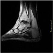
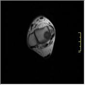
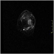
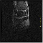
Osteochondritis dissecans of the talus


Download
Info
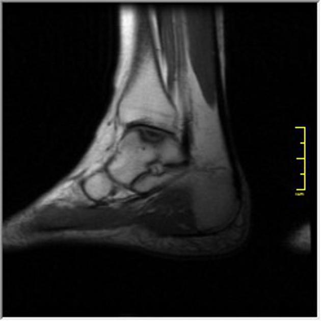
An osteochondral bone fragment is seen at the superomedial aspect of the talar dome. It shows diffuse sclerosis and is surrounded by a sclerotic line of its cavity. A linear high T2 and STIR signal (rim sign) is surrounding the osteochondral fragment suggesting fragment separation. No detachment is seen.
Case Discussion
Features of stage 3 osteochondritis dissecans involving superomedial aspect of the talar dome.


 Unable to process the form. Check for errors and try again.