
Presentation
Flank pain
Patient Data
Age: 18 years
Gender: Female


Download
Info
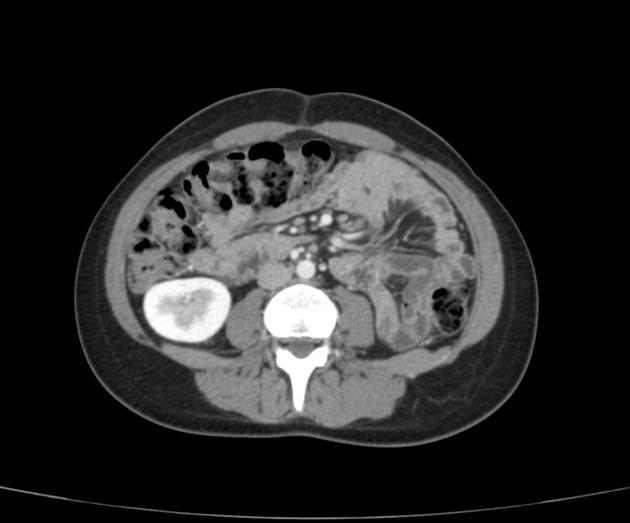
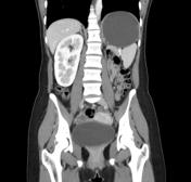
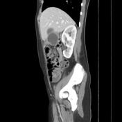
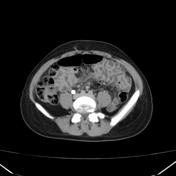
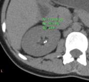
CT of the abdomen demonstrates a solitary right kidney, with physiological hypertrophy. The morphology of the right kidney is normal and shows normal concentration and excretion of contrast media.
Right renal upper calyceal stone is noted.
Case Discussion
The solitary kidney, apart from compensatory hypertrophy usually is present from birth. It is usually normal, and its discovery is feasible when the radiographic examination should be performed for a kidney condition. It is then discovered that it is a solitary kidney.


 Unable to process the form. Check for errors and try again.