
Presentation
Swelling at the back of long duration
Patient Data
Age: 17 years old
Gender: Male
From the case:
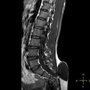
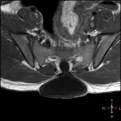
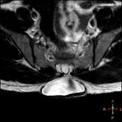
Myelomeningocele


Download
Info

S3 spina bifida with a sac like structure seen connected to the spinal canal and filled with CSF. Flow artifact could be seen at the ostium of this cystic lesion in T2 WI. The cord is in normal position.
Case Discussion
Spinal dysraphism associated with myelomeningocele.
Differential diagnosis - post operative pseudomeningocele
Related article


 Unable to process the form. Check for errors and try again.