Bosniak renal cyst - type II

Case contributed by Andrew Dixon
Diagnosis certain

Triphasic Renal CT

ct
This study is a stack
Axial
non-contrast
This study is a stack
Axial renal
cortical phase
This study is a stack
Coronal renal
cortical phase
This study is a stack
Axial renal
parenchymal phase
This study is a stack
Coronal renal
parenchymal phase
This study is a stack
Axial renal
excretory phase
Download
Info

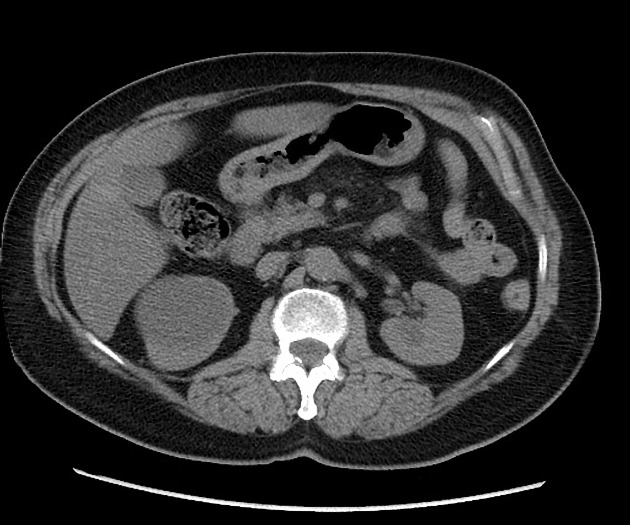
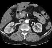
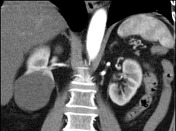
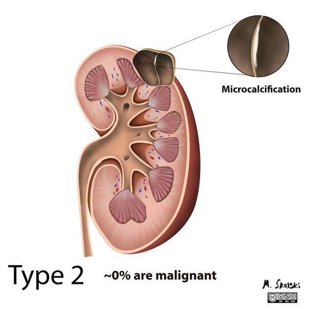
Left upper pole renal cyst with a single thin focus of wall calcification. Right lower pole renal cyst with a single thin septation. Both cysts fall into the 'minimally complex' Bosniak II category and can be confidently said to be benign without follow-up imaging required.  

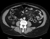
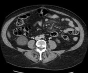
Incidental findings include mesenteric panniculitis, mildly fatty liver and uterine fibroids. 

Illustration
Download
Info

Illustrative example of a Type 2 cyst.

How to use cases

You can use Radiopaedia cases in a variety of ways to help you learn and teach.

Creating your own cases is easy.

:

Updating… Please wait.

 Unable to process the form. Check for errors and try again.

 Thank you for updating your details.