
Presentation
Neck pain with bilateral upper limbs parasthesia.
Patient Data
Age: 55 Years
Gender: Female
From the case:
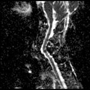
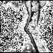
Intradural extramedullary arachnoid cyst - cervical spine

Download
Info

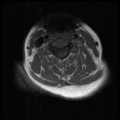
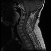
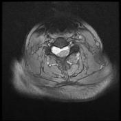
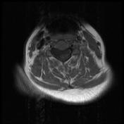
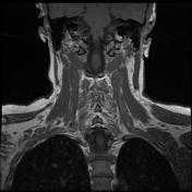
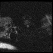
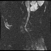
- a lobulated creeping right-sided intra-spinal
intradural extra-medullary predominantly cystic lesion is seen, extending from
C2/C3 till’ C7/D1 level.
- it shows heterogenous; predominantly hypointense T1 and hyperintense T2 and GRE signal intensity.
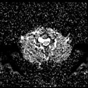
- the lesion shows facilitated diffusion with no fluid restriction (low signal on DWI and high signal on ADC ).
- the lesion shows no evident post-contrast enhancement.
- the lesion measures about 5.5 X 2.5 X 1.3 cm in its main CC, and axial diameters respectively. The lesion is seen compressing and displacing the spinal cord posteriorly and to the left side.
- normal cervical cord and cervico-medullary junction with no cord syrinx or abnormal signal intensity.
- fairly good vertebral alignment.
- no posterior disc lesion.
- no marrow signal abnormality.
- no para-spinal masses.
Conclusion
A lobulated and creeping intra-spinal intradural extra-medullary non-enhancing cystic lesion, mostly arachnoid rather than epidermoid cyst.


 Unable to process the form. Check for errors and try again.