
Presentation
Right sided SNHL, headache and tinnitus.
Patient Data


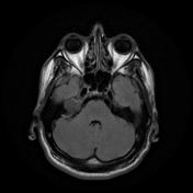

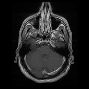
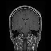
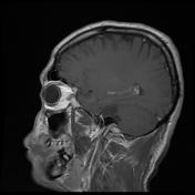
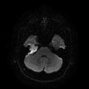

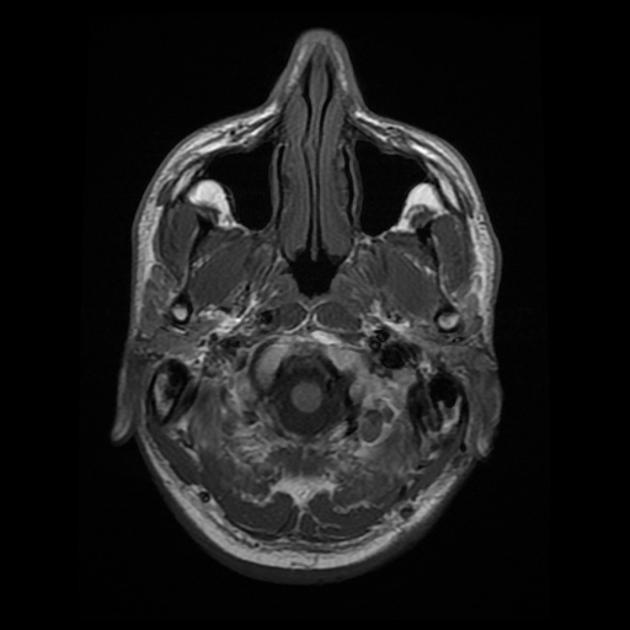
A right petrous apex well-defined lobulated border S.O. lesion is seen. It measures 3.2 X 1.8 X 1.9 cm in its main axial and CC diameters respectively. The lesion shows hypointense T1 and bright T2 signal with incomplete nullification of its signal in the FLAIR sequence. The lesion is hyperintense in DWI series. It is slightly hypointense to isointense to the brain in ADC map. The lesion shows no significant post-contrast enhancement but for subtle / if any marginal enhancement. The lesion is seen mildly compressing the right internal auditory canal the right facial and vestibule-cochlear nerves as well as the right cerebello-pontine angle and abutting the right inferior temporal lobe.
Case Discussion
Right petrous apex mass lesion - which most likely represents a petrous apex cholesteatoma.
A cholesteatoma will not usually attunuate on FLAIR imaging, however partial attenuation, as seen here, is not uncommon.


 Unable to process the form. Check for errors and try again.