
Presentation
Circus performer with several months of increasing dorsal wrist pain.
Patient Data


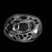
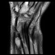
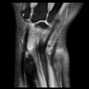
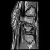
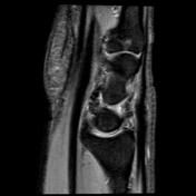
There is a high signal within the extensor pollicis longus (EPL) tendon just distal to Lister's tubercle as it crosses over the second extensor tendons (ECRB and ECRL) and is flattened by the overlying extensor retinaculum. There is localized tenosynovitis around this area and a small amount of increased fluid is present within the EPL and ECRL tendon sheaths distally. This appearance is consistent with distal intersection syndrome. There is also thickening and hyperintensity of the dorsal wrist capsule which is likely due to repetitive sprain. A ganglion / effusion is noted at the triquetral-pisiform articulation. A linear cleft is seen within the proximal (membranous) portion of the scapholunate ligament with the volar and dorsal bands intact. There is a loss of cartilage at the scaphoid-trapezoid articulation.
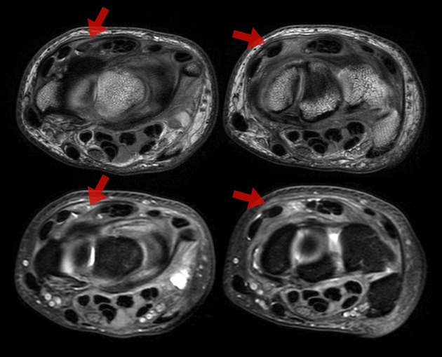
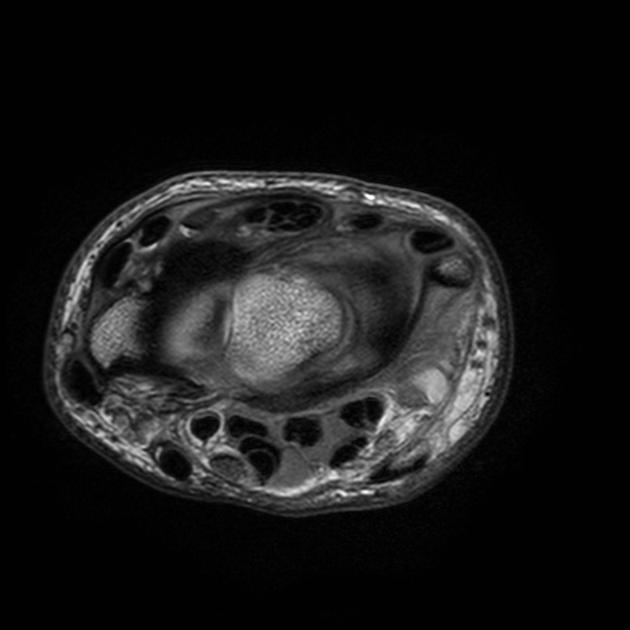
Top row - axial PD images showing loss of the normal low signal with EPL just before and as it crosses the second extensor compartment tendons.
Bottom row - axial PD fat sat images at the same level with the EPL virtually invisible amongst the surrounding tenosynovitis.
Case Discussion
Features of distal intersection syndrome (EPL tenosynovitis) in the setting of overuse.


 Unable to process the form. Check for errors and try again.