
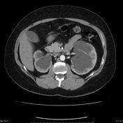

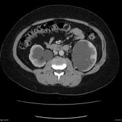

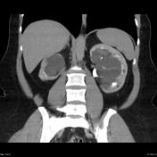
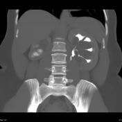
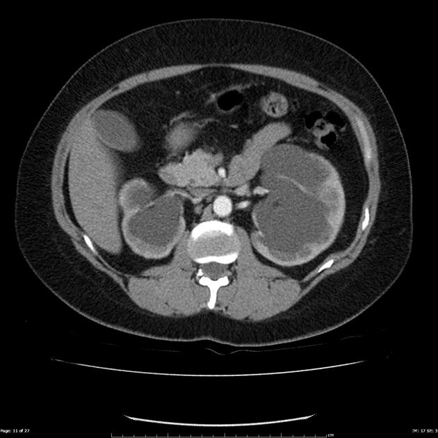
A three-phase CT study of the kidneys has been performed (corticomedullary, nephrographic and excretory) phases. The excretory phase equates to a CT-IVU, typically undertaken at 8-10 minutes following IV contrast administration.
The pelvis of both kidneys are expanded by water density spaces. The right kidney is atrophic with a reduced bipolar diameter (BPD). The left is larger. The cortex of both kidneys enhances normally.
On the delayed excretory phase images, the kidneys behavior differently. On the right contrast pools dependently within a distended renal pelvis, consistent with a PUJ obstruction. On the left the calyces are outlined, compressed by numerous para-pelvic cysts (which do not contain contrast).
Case Discussion
This case elegantly demonstrates how difficult it can be to distinguish a dilated renal pelvis from parapelvic cysts without a delayed phase.


 Unable to process the form. Check for errors and try again.