
Presentation
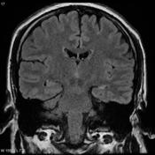
Temporal lobe epilepsy
Patient Data
Gender: Male
From the case:
Mesial temporal sclerosis


Download
Info
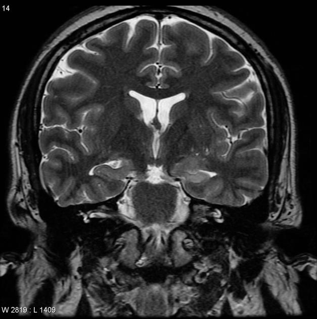
Reduced volume of the left hippocampus compared to the right, with an area of subtle hyperintense signal on FLAIR sequences. This represents neuronal loss and gliosis consistent with left sided mesial temporal sclerosis.
Case Discussion
The diagnosis was confirmed with histopathological analysis.


 Unable to process the form. Check for errors and try again.