
Presentation
This patient felt a lump in her left breast. She attended the triple assessment clinic.
Patient Data
Age: 30 years
Gender: Female

Download
Info

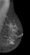
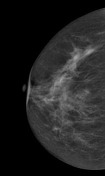
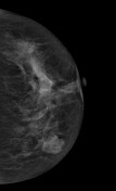
Both breasts are composed of scattered fibroglandular tissue. In the left breast at the 8 o'clock position, there is a lobular circumscribed high density mass without calcifications. No lymph nodes seen.

From the case:
Fibroadenoma of the breast
Download
Info
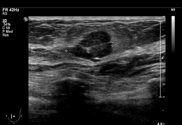
Corresponding to the mammogram, there is a lobulated sharply demarcated mass in the left breast at the 8 o'clock position with a thin echogenic rim (pseudocapsule). The mass is generally hypoechogenic.
Case Discussion
In this case, there are no radiological or clinical red flags and the lesion has typical features of a fibroadenoma. No biopsy required; clinical follow up only. BIRAD 2.


 Unable to process the form. Check for errors and try again.