
Presentation
Elite dancer with two weeks of left gluteal region pain.
Patient Data

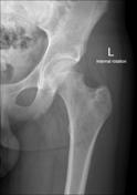
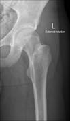
No definite radiographic abnormality.
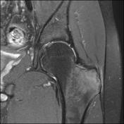

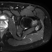
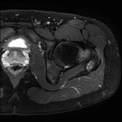
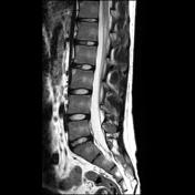
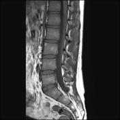

Marrow edema within the greater trochanter and gluteus medius and minimus peritendinous edema. Left sacroiliitis is also seen. Incidental low grade chondoid lesion within the subtrochanteric region. Lumbar spine MRI reveals "shiny corners" at the anterior corners of several of the lumbar vertebral bodies, an early feature of ankylosing spondylitis.

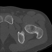
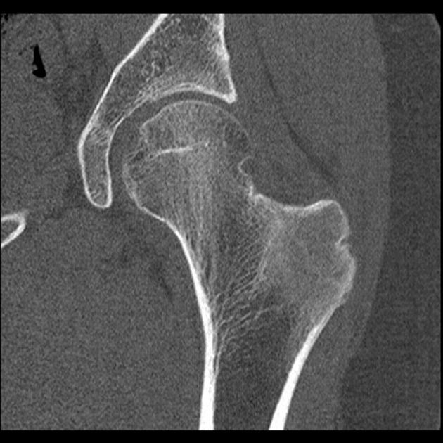
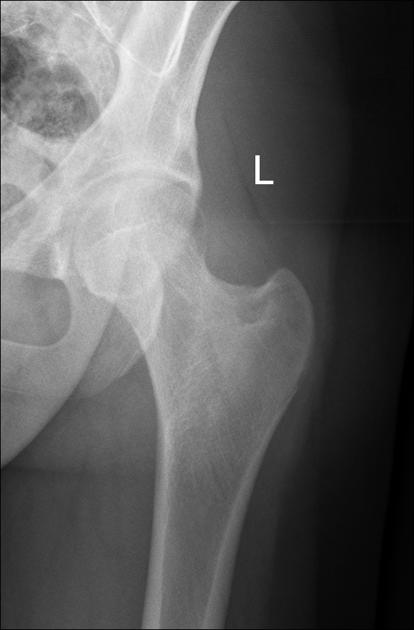
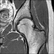
Subtle new bone formation at the gluteal enthesis (enthestitis) is seen at the greater trochanter. No other CT abnormality.
Case Discussion
A case of ankylosing spondylitis presenting as gluteal enthesitis. This case emphasizes the importance of looking at the sacroiliac joints on MRI of the hip for signs of sacroiliitis and the importance of recognizing "shiny corners" as an early feature of ankylosing spondylitis in the lumbar spine on MRI.


 Unable to process the form. Check for errors and try again.