
Presentation
History of pain at knee during flexion, with a palpable swelling at posteriomedial aspect of knee. Clinically thought to be a soft tissue tumor.
Patient Data
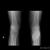

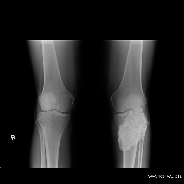
A fairly large lobulated calcified cauliflower like lesion , almost tear shaped seen at posteromedial soft tissue of knee joint.
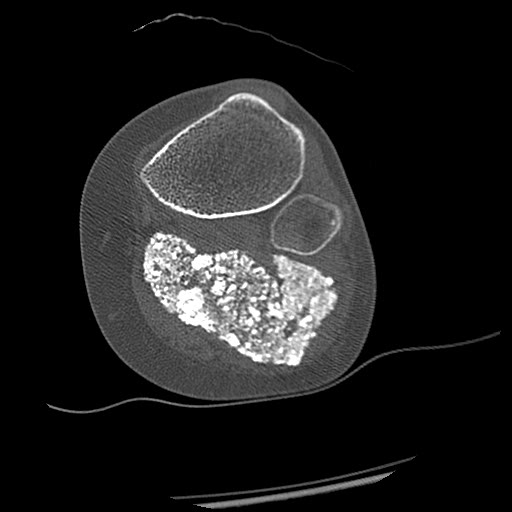
Bone algorithm CT showed the same lesion as xray, added no extra information, except that this calcified mass was splaying the muscles at the posteromedial aspect of knee and not expanding them, hence ruling out an intramuscular tumor. There are areas of discontinuity at the peripheral margins not seen on Xray , but with no obvious soft tissue component.
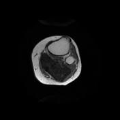

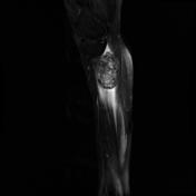
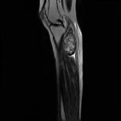

A large lobulated lesion, now clearly seen to be lying in the enlarged redundant popliteal recess of knee joint, splaying the medial head of gastrocnemius and semimembranosus. The internal matrix is heterogenous predominantly mottled hyperintense signal on T2 weighted and hypointense signal on T1 weighted sequences, margins are well defined, and there's no intraosseous or intramuscular extension suggesting a benign calcified mass.
The radiological diagnosis was solitary giant intrasynovial loose body of popliteal recess of knee.
Case Discussion
This lesion was surgically resected. At surgery it was pedunculated with frail septae connecting it to the synovium of the posterior popliteal recess of knee.
Histopathology showed islands of dystrophic calcification with some cartilaginous tissue at periphery, a diagnosis consistent with a giant solitary loose body of popliteal fossa. Very few such solitary giant loose bodies have been reported in literature. This one measured 7 x 4.7 x 6 cm.


 Unable to process the form. Check for errors and try again.