
Presentation
Seizures
Patient Data
Age: 50 years
Gender: Male
From the case:
Polymicrogyria


Download
Info
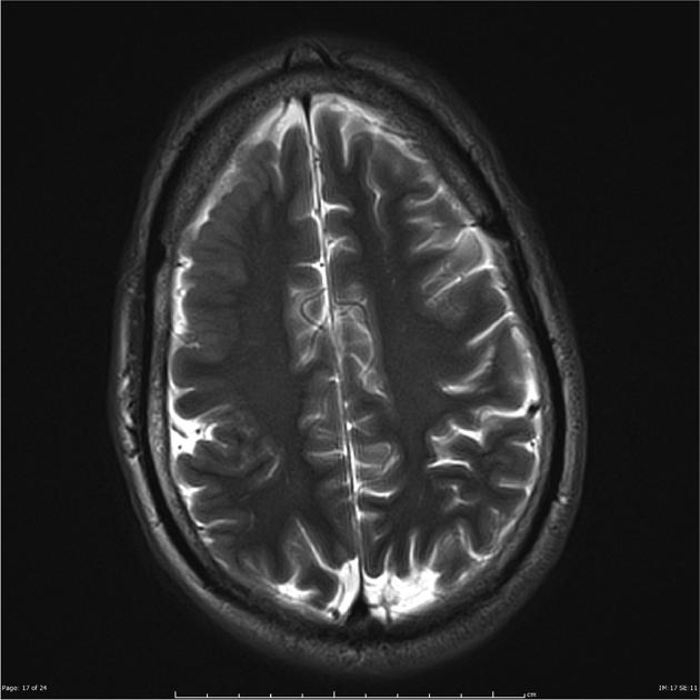
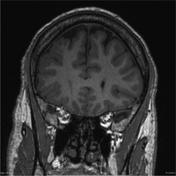
The right cerebral hemisphere is markedly abnormal. It is slightly smaller than the left side and demonstrates abnormal sulcation and cortex. The abnormality is most marked in the superolateral right frontal and parietal lobes where there is mild cortical thickening and increased gyrus. The abnormality extends into the posterior temporal region. There is a mildly prominent sulcus in the superior parietal region. The cortex in the right insular region is thickened, likely dysplastic.
Case Discussion
Brain MRI findings compatible with polymicrogyria are most marked in the right frontal and parietal lobes.


 Unable to process the form. Check for errors and try again.