
- Note: This case has been tagged as "legacy" as it no longer meets image preparation and/or other case publication guidelines.
From the case:
Hypertrophic olivary degeneration

Download
Info
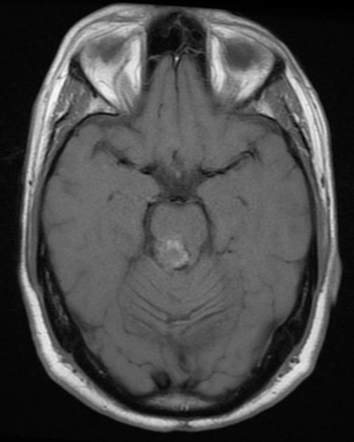
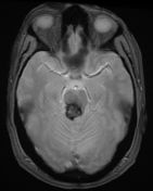
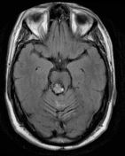
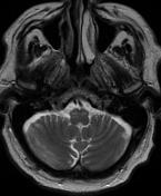
Cavernous malformation of midbrain resulting in bilateral (right > left) increased T2 signal and slight mass effect of the inferior olivary nuclei, suggesting the diagnosis of hypertrophic olivary degeneration.
Case Discussion
This case is subtle and may be unilateral (right only), although increased T2 signal is seen in both olives. Palatal myoclonus would help confirm the diagnosis.


 Unable to process the form. Check for errors and try again.