
Presentation
Sudden onset of headache, hypothyrodism, low TSH and visual defect.
Patient Data
Note: This case has been tagged as "legacy" as it no longer meets image preparation and/or other case publication guidelines.
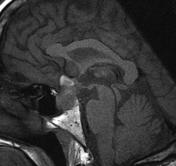
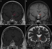
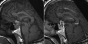
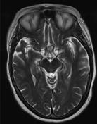

Sagittal T1 images demonstrate a heterogenous sella mass with suprasellar extension leading to impression of the optic chiasm and infundibular recess of the third ventricle. Areas of high T1 signal are seen in the superior portion of the mass which may represent hemorrhage/methemoglobin. This area of high signal intensity fails to suppress on the coronal T1 FS. Coronal and axial T2 images confirm intralesional hemorrhage with peripheral hemosiderin staining and a central region of mixed signal intensity representing different stages of blood product degradation. The mass demonstrates a peripheral and nodular pattern of enhancement. Presella pneumatization of the sphenoid sinus. Overall imaging pattern is most in keeping with pituitary macroadenoma with intratumoral hemorrhage.
Case Discussion
Pituitary apoplexy is characterized by clinical presentation of headache, visual disturbance and endocrine disturbance. Ischemia and hemorrhage of a pre-existing macroadenoma is the most common cause of pituitary apoplexy.


 Unable to process the form. Check for errors and try again.