
Presentation
Seizures.
Patient Data
Age: 42
Gender: Male
From the case:
Mesial temporal sclerosis
Download
Info
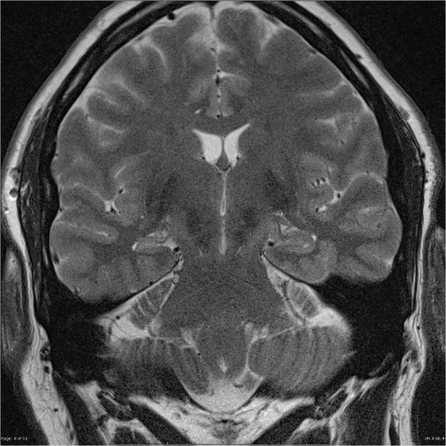
The right hippocampus is markedly smaller than the left with loss of the normal internal architecture and diffuse T2 high signal which involves the body and tail. The amygdala is unremarkable. The remainder of the temporal lobes are symmetrical. No evidence of associated neuronal migration abnormalities. The remainder of the brain is unremarkable with no evidence of restricted diffusion or regional susceptibility induced signal loss.
Conclusion: The imaging findings are consistent with mesial temporal sclerosis.


 Unable to process the form. Check for errors and try again.