Presentation
CT scan to elucidate multiple echogenic nodules on ultrasound study.
Patient Data
Age: 65 years
Gender: Male
![](https://prod-images-static.radiopaedia.org/images/5330404/f0f3b179ecb3fac83e275fc8271b821659bbede2210cd97806c9e1a9dd45dbc5_thumb.jpeg)
![This study is a stack](/packs/stack-YQKLCKBI.gif)
![](https://prod-images-static.radiopaedia.org/images/5329782/4a2a04419a8a4186666bf28d49c10d8ab4f91b45d63e129aaaa1a8d5cfa1a74e_thumb.jpeg)
![This study is a stack](/packs/stack-YQKLCKBI.gif)
![](https://prod-images-static.radiopaedia.org/images/5330196/bf162be4c3de594cd3e58dc521b7eccd243c293a6d7a5af3dde2d8706958c35f_thumb.jpeg)
![This study is a stack](/packs/stack-YQKLCKBI.gif)
![](https://prod-images-static.radiopaedia.org/images/5330067/aa4319898be5b56cd683993046c8871918fdacab87911d314de71d149941e34c_thumb.jpeg)
![This study is a stack](/packs/stack-YQKLCKBI.gif)
Download
Info
![](https://prod-images-static.radiopaedia.org/images/5330404/f0f3b179ecb3fac83e275fc8271b821659bbede2210cd97806c9e1a9dd45dbc5_big_gallery.jpeg)
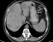
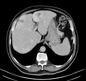
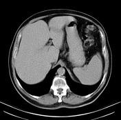
Multiple hypoattenuating lesions in the liver that show progressive, peripheral and globular enhancement, becoming homogeneous to hepatic parenchyma in the equilibrium phase.
Case Discussion
A hepatic hemangioma is a benign hypervascular liver lesion. It is the most common benign tumor of the liver.