
Presentation
Diabetic patient presenting with left flank pain and fever.
Patient Data

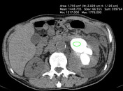
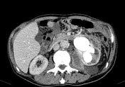


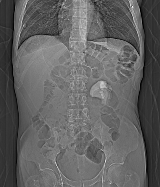
Large staghorn calculi in the left kidney collecting system. Renal calyces are dilated giving a multiloculated appearance. There is also a retroperitoneal collection that extends along the left iliac and psoas muscle.
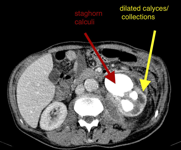
Large staghorn calculi in the left kidney collecting system. Renal calyces are dilated giving a multloculated appearance.
Case Discussion
Xanthogranulomatous pyelonephritis is a chronic destructive granulomatous process of renal parenchyma in association with long-term urinary tract obstruction and infection.
Clinical presentation includes flank or abdominal pain, lower urinary tract symptoms, fever, palpable mass, hematuria and weight loss.
This case shows a xanthogranulomatous pyelonephritis associated with staghorn calculus and complicated with extensive retroperitoneal abscess.


 Unable to process the form. Check for errors and try again.