
Presentation
History of back pain for 4 weeks. Presents with acute worsening without trauma.
Patient Data
Age: 60 years
From the case:
Pathological vertebral crush fracture - plasmacytoma

Show annotations
Download
Info
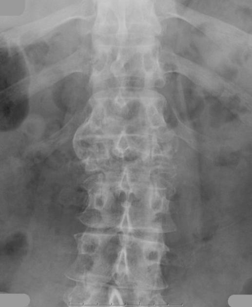
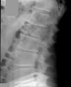
A pathological compression fracture of L1 is seen on plain films.
From the case:
Pathological vertebral crush fracture - plasmacytoma

Show annotations
Download
Info

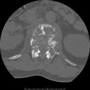
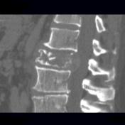
CT axial image through L1 and sagittal reconstructions show a heterogenous lytic lesion of the vertebral body and posterior elements.
Case Discussion
The diagnosis of plasmacytoma was suggested by percutaneous needle biopsy and confirmed at surgery when a rib strut graft and plate were placed between T12 and L1.


 Unable to process the form. Check for errors and try again.