
Presentation
Headache, nausea, vomiting, blurred vision and seizure.
Patient Data


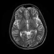
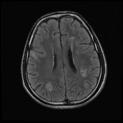

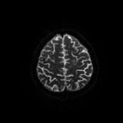
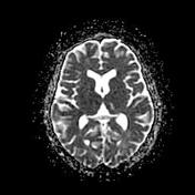
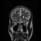

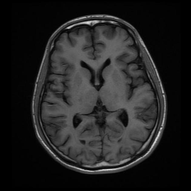
Multiple ring enhancing lesions which are isointense to gray matter on T1, hypointense on T2 and FLAIR with surrounding vasogenic edema seen involving bilateral precentral gyri, bilateral centrum semiovale and corona radiate, left angular gyrus, bilateral middle frontal gyri, right superior frontal gyrus, left inferior frontal gyrus, left superior and middle temporal gyrus, inferior cerebral peduncle and mid brain.
No blooming on SWI and no hyperintensity on DWI or ADC is noted.
Imaging features and clinical profile of the patient are suggestive of multiple tubercular granulomas.
Case Discussion
This patient is a known case of pulmonary tuberculosis, who has not completed the treatment.
Intracranial tuberculomas are space occupying lesions that result from hematogenous spread of primary infection from a distant focus. Histologically the central iso or hypointense area contains caseous necrosis surrounded by a capsule (enhancing) made up of fibroblasts, epithelioid cells, Langhans giant cells and lymphocytes.


 Unable to process the form. Check for errors and try again.