Presentation
Right flank pain for the past 1 month. USG revealed right sided hydronephrosis. A mid ureteric calculus was suspected.
Patient Data


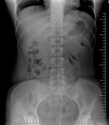
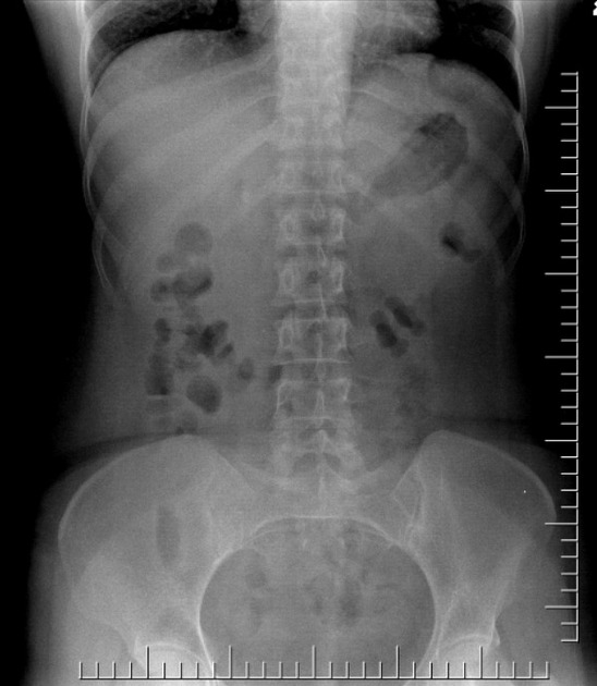
Spot films taken during an IVP
The 1st image is a scout film which revealed no calculus.
The 2nd film is a radiograph taken after 10 mins of injection of contrast. In this we see a dilated renal pelvis due to kink in the ureter at the level of L3 vertebra.
Another point of note is the pelvis on the right side is extrarenal in nature.
Case Discussion
Case of ureteric kink and right sided extrarenal pelvis with no calculus.
These are the causes of dilatation of the pelvi-calyceal system which has been misinterpreted on the ultrasound as a dilatation due to calculus disease.


 Unable to process the form. Check for errors and try again.