
Presentation
Two weeks of headache, low grade fever and Wernicke aphasia. Previews history: recurrent ear infections. WBC within normal limits and negative HIV test.
Patient Data
Age: 32 year old
Gender: Female


Download
Info
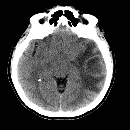
Round intra-axial lesion with vasogenic edema. Post contrast shows a thin and smooth wall. Coronal and sagital reformats shows no mural nodule.
From the case:
Brain abscess


Download
Info
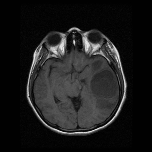
Round intra-axial lesion with vasogenic edema that have a hyper intense thin-wall in T1 and hypo intense wall at T2. Post gadolinium shows rim-enhancement.
Case Discussion
The CT and MRI findings are compatible with pyogenic brain abscess in late capsular stage.


 Unable to process the form. Check for errors and try again.